
Notably, it is among the few private MRI clinics in Bucharest to have a 3 Tesla MRI machine (GE Signa HDxt), which is known for its superior image quality and diagnostic accuracy. This advanced technology is instrumental in addressing a wide range of medical conditions with precision.
Additionally, the clinic's commitment to quality is underscored by its accreditation and certification according to ISO 9001:2008 standards. This certification reflects the clinic's adherence to international standards in healthcare service quality, ensuring that patients receive the best care in a safe and professionally managed environment.
As medical technology evolves, Biomed Scan Clinic is likely to continue integrating cutting-edge advancements, maintaining its position as a pivotal player in Bucharest's medical imaging arena.
MRI vs. CT Scans: While both MRI and CT scans are used for diagnostic imaging, MRI does not use ionizing radiation like CT scans. MRI provides better contrast in soft tissues, making it ideal for imaging organs, soft tissues, and the central nervous system.
General MRI Examinations: These include cerebral MRI, lumbar MRI, abdominal MRI, and more. Specialized exams, such as Angio RMN cerebral, are also available. The clinic offers a comprehensive range of MRI services to suit various diagnostic needs.
The clinic prides itself on its advanced MRI technology. Equipped with high-end MRI systems from renowned manufacturers such as Siemens and General Electric, Biomed Scan offers both 1.5 Tesla and 3 Tesla imaging capabilities.
Pacemakers and Implantable Devices: Patients with pacemakers or certain other implantable electronic devices may not be eligible for an MRI.
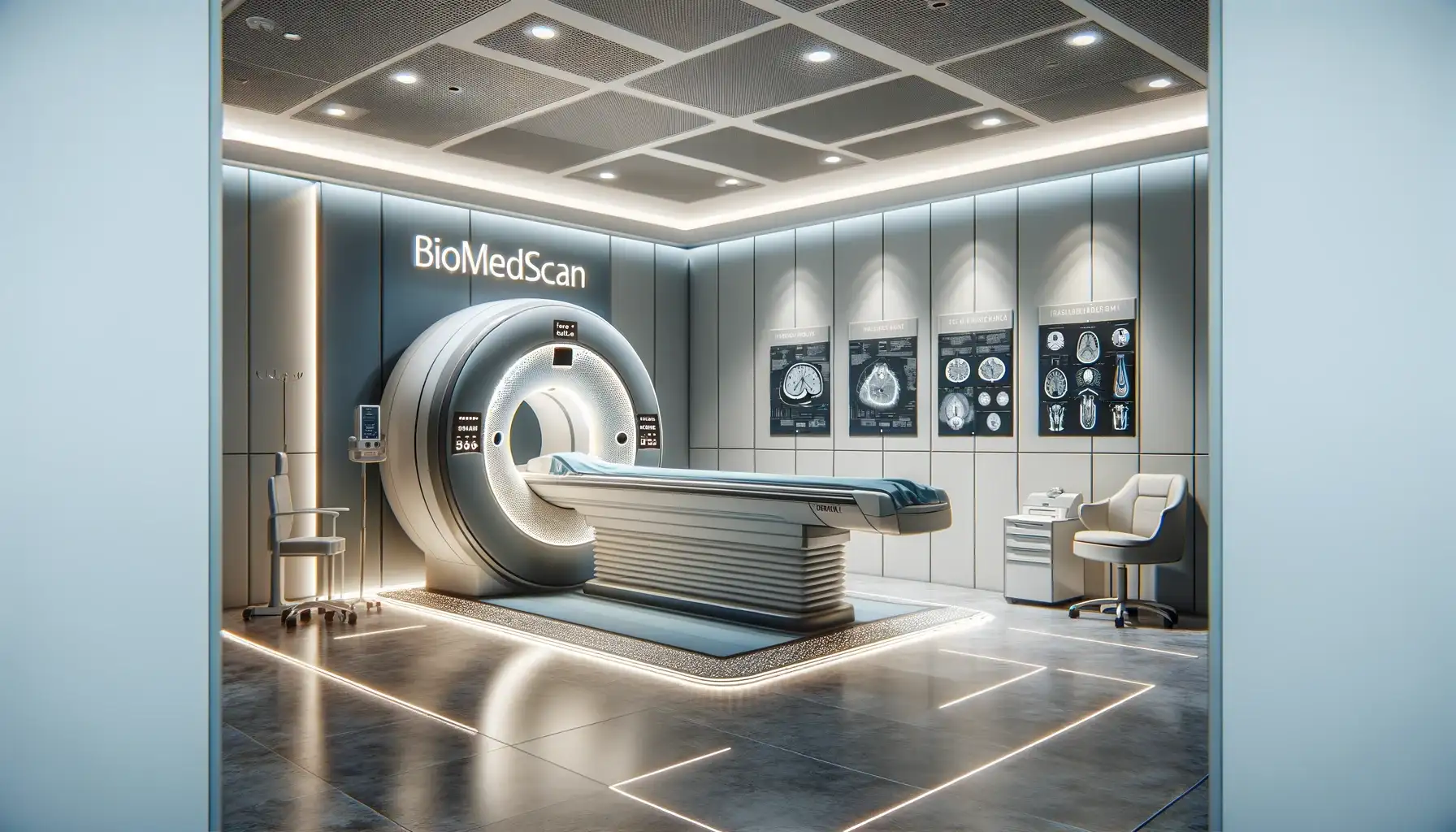
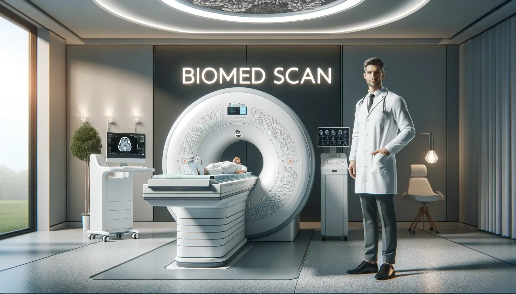
Facilities and Equipment
Communication: Throughout the examination, patients can communicate with the medical practitioner or MRI operator through a microphone.
History and Location
Pregnancy: MRI scans are typically avoided during the first trimester of pregnancy, and pregnant patients should consult their doctor before undergoing an MRI.
This includes, but is not limited to, neurological disorders (such as brain tumors, multiple sclerosis, and epilepsy), orthopedic conditions (like fractures and joint problems), oncological cases (such as breast, prostate, and liver tumors), and various cardiovascular and abdominal organ diseases.
MRI Services Offered at Biomed Scan Clinic
Unique Advantages of MRI:
The clinic’s journey, from its inception to becoming a leading MRI service provider in Bucharest, reflects its dedication to healthcare excellence. With a focus on patient-centric services, Biomed Scan has equipped itself with cutting-edge MRI machines and has cultivated a team of over 24 experienced radiologists and MRI operators, ensuring that each examination is conducted with the utmost precision and care.
Screening for Metal Objects: Due to the strong magnetic field, patients are screened for any metal objects or implants before an MRI scan.
Join us as we navigate through the intricacies of RMN Bucuresti services at Biomed Scan Clinic, unveiling a world where technology and healthcare converge to provide unparalleled diagnostic accuracy and patient care.
Looking ahead, the future of MRI technology in Bucharest, with Biomed Scan Clinic at the forefront, appears promising. The non-invasive, radiation-free nature of MRI makes it an increasingly preferred diagnostic tool, particularly beneficial for young patients, pregnant women, and those requiring repeated screenings.
MRI and X-rays/Ultrasound: MRI offers more detailed images compared to X-rays and ultrasound. X-rays are typically used for imaging bones and joints, while ultrasound is used for soft tissue and organ imaging. MRI, however, provides a more comprehensive view of both soft and hard tissues.
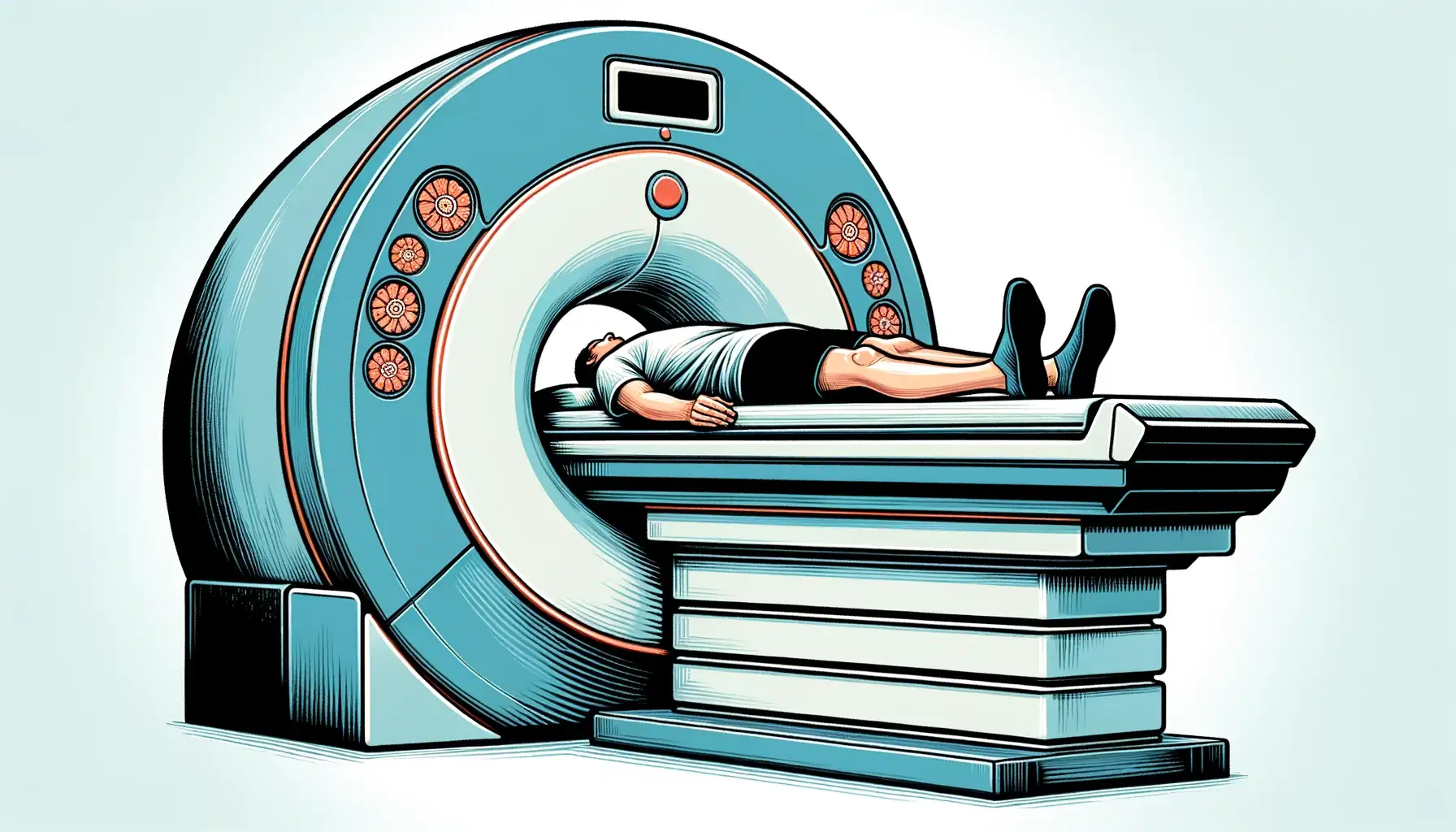
MRI Examination Process:
MRI sequences are sets of parameters that dictate how the MRI machine will operate to create images. Spin echo and gradient echo are types of sequences that manipulate the protons' behavior in the body, providing different types of image contrast and resolution.